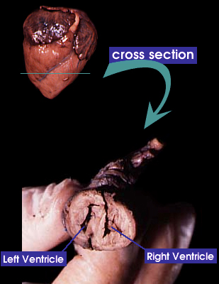
Pig heart cross section
 This image shows a cross section of the fetal pig heart, created by cutting through the ventricles of the heart at the level indicated.
The ventricles are separated by the interventricular septum. The atria are also separate from one another, so that the de-oxygenated blood of the right chambers is kept separate from the oxygenated blood of the left chambers.
In the mammalian fetus, the left and right ventricular walls are about equal in thickness. Following birth the left ventricular wall gradually becomes thicker because the left ventricle has to pump blood out to the systemic circuit. This is very long (with high resistance) and requires blood to be pumped at a higher pressure via thicker cardiac muscle.
Next: Cow heart
Back to: The circulatory system