Diaphragm
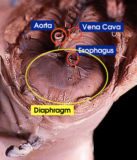
 This photo shows the diaphragm at the caudal end of the opened thoracic cavity -- the view is from the head of the pig looking caudally (the ventral portion of the rib cage and thoracic organs have been removed; the dorsal portion of the rib cage is visible at the top of the image).
The diaphragm (in yellow oval) is visible as a sheet of skeletal muscle, which separates the thoracic and abdominal cavities.
To get air into the lungs (inspiration), the rib cage expands and the diaphragm contracts to enlarge the thoracic cavity, generating a negative pressure within the lungs, causing air to flow in.
To eject air from the lungs (expiration), the diaphragm and rib muscles (e.g., intercostal muscles) relax, reducing the thoracic cavity volume and generating a positive pressure that pushes air out.
Back to: Respiratory system overview